


Skin lesion removal

Skin and soft tissue lesions are very common. Most commonly they are benign lesions; melanocytic nevi, lipomas, atheromas, fibromas or warts (f.e. verruca). Despite their aesthetic aspect, lesions are subject to re-occuring inflammations and infections. The most serious danger and cancerous lesions, which can resemble benign lesions or develop from them (Malignant transformation, malignancy).Skin and soft tissue lesions are very common. Most commonly they are benign lesions; melanocytic nevi, lipomas, atheromas, fibromas or warts (f.e. verruca). Despite their aesthetic aspect, lesions are subject to re-occuring inflammations and infections. The most serious danger and cancerous lesions, which can resemble benign lesions or develop from them (Malignant transformation, malignancy).
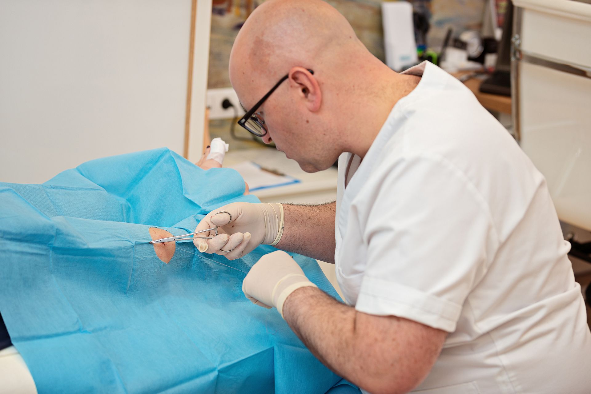
What does lesion removal look like?
Zabieg wycięcia zmian skórnych przebiega w znieczuleniu miejscowym. Po wycięciu rana zostaje zszyta tak, aby pozostawić jak najmniej widoczną bliznę. Następnie na ranę zakłada się jałowy opatrunek i pacjent może wrócić do domu. Skin lesions removals are done under the local anaesthesia After the removal the wound is stitched to leave an almost invisible mark. Then a sterile dressing is applied to the wound and the patient can go home.
Recommendations after treatment
After the procedure the wound has to be disinfected daily and the dressing has to be changed every day. You can do it yourself at home or come to our centre upon making an appointment with a qualified nurse or a doctor. A shower can be taken already after 3-4 days. If non-absorbable sutures have been left, they should be removed after 7-10 days. Sometimes absorbable suture is used, which does not have to be removed.
What happens with the excised skin?
After the lesion has been excised, it will be sent for histopathological examination, which will determine the character of the excised skin. It impacts further steps. The wait time for the results is approximately 4-5 weeks.
In certain cases, in order to confirm the diagnosis, in addition to macroscopic and microscopic evaluation, additional tests need to be performed. The type and number of additional tests are decided by the Department of Pathology which supervises the diagnostic procedures. Additional tests may include cytology, immunochemistry, EBV testing and specialist consults.
Consult our specialists:
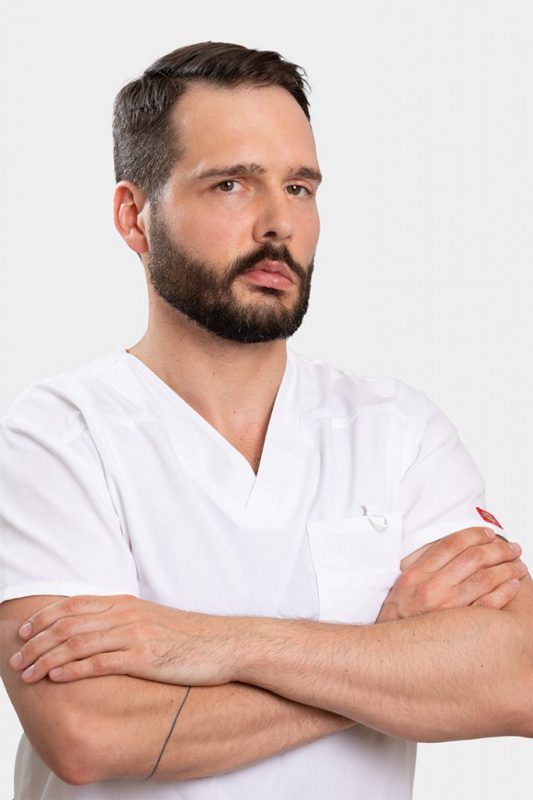
Kamil Dąbrowski, M.D.
General surgery and surgical oncology specialist, certified robotic surgeon
Book appointment