


Pilonidal disease (hair cyst) – is it possible for the disease to pass by itself?
Pilonidal disease is a chronic skin infection which typically occurs as a cyst in the crease of the buttocks near the tailbone and is characterized by periodic inflammations. Its characteristic feature is the hair growing in the cleft – hence the name, cyst (sinus) or pilonid (from the Latin words pilus – hair and nidus – nest).
Symptoms
The location of the cyst causes the bacteria to multiply inside. This is causing inflammation of the surrounding tissue, which in turn can cause pain, fever and fluid drainage from affected area with an unpleasant smell. The inflammations become more frequent and the cyst becomes more extensive.
Effective treatments of a pilonid cyst
Unfortunately, there is no effective home treatment for cysts except for painkillers and anti-inflammatory drugs. In the event of a fever, antibiotics are needed. There has to be a surgical intervention in case of an abscess. The surgery involves then cutting the wall of a cyst, evacuating the pus, flushing and draining its light. After the inflammation subsides, pain decreases and the wound heals. However, this does not mean getting rid of the problem, because the entire anatomical structure is still under the skin.
Surgical treatment is the only effective way to treat a hair cyst. Traditional surgery involves extensive cyst excision requiring a stay at the hospital, because of an extensive postoperative wound that heals for a long time. Due to the frequent complications of wound healing, most surgeons leave it open, which extends healing time up to several months.
Laser ablation of a pilonid cyst
Currently, laser cyst ablation is one of the most effective and least invasive methods of surgical treatment. The advantage is its very high efficiency, and can also be compared to the best classical methods. There is a small, quickly healing wound and because it is an outpatient type of surgery, the patient can immediately go home. By putting the optical fiber inside the cyst and destroying its walls using laser energy, there is no need to cut it, which significantly reduces the scope of surgery.
Laser ablation of the hair cyst (pilonidal sinus laser therapy – SiLaT) is a procedure that can be performed either by giving local anesthesia, or by administering anesthesia to the canal of the spinal cord or by not using anesthesia at all. The procedure itself takes about 20-30 minutes and the patient can leave the clinic right after the treatment. The minimal postoperative wound does not interfere with normal lifestyle and does not cause significant pain. The effectiveness of the method appearing in clinical trials is estimated at 92-95%, while the most common simple excision of the lesion, has an efficiency of 85%. It should also be noted that among all available methods, SiLaT leaves the best cosmetic effect.
Author: VenoMedica
All rights reserved.
Author: venomedica
Date: 2020-07-04
How to treat varicose veins and avoiding complications?
Varicose veins are a symptom of venous insufficiency. They are most often caused by insufficient valves in the main trunks of the superficial veins – in the great and small saphenous veins. Venous insufficiency is usually genetic. Incorrect structure of the vessel walls causes a loss of elasticity and a tendency to stretch them. The choice of varicose vein treatment method has an impact on its effectiveness, safety and cosmetic effect.
The primary goal of any treatment is to block the back flow of the blood to the venous system, which causes the following:
- that venous pressure increases,
- blood stasis in widened blood vessels,
- swelling of the subcutaneous tissue,
- the inflammatory changes,
- the varicose vein thrombosis,
- discoloration,
- skin ulcers.
Varicose veins treatment methods can be divided into two groups:
CLASSIC SURGICAL METHODS:
- stripping
- miniflebectomy
MINIMALLY INVASIVE ENDOVASCULAR METHODS:
- thermal
- chemicale
- mechanical and chemical
Surgical methods
The first group includes the oldest surgical methods – stripping and miniflebectomy. This kind of technique was used for the first time in 1905 by William Wayne Babcock, where he had to remove the saphenous vein part using a flexible, special probe. Due to the small number of relapses and a relatively low percentage of serious complications, this method has been carried out until today. It is complemented by so-called miniflebectomy, i.e. a surgical operation to remove varicose veins using punctures or small skin incisions. The surgery is most often performed under either spinal anesthesia (an anesthetic injected into the central canal of the spinal cord) or general anesthesia.
There can be, however, one very serious complication using the stripping method. The deep vein or arterial system can be severly damaged as well as its nerve trunks. This can lead to permanent paralysis of muscle groups or sudden numbness affecting a certain skin area. Furthermore, postoperative wound infection occurs in 2-3% of the cases. Paradoxically, after classical surgery, there may be an increase in limb swelling caused by impairment of lymph nodes and lymphatic vessels.
While the long-term results of classic operations are quite good, in the postoperative period the pain is significantly worsened. This is caused by extensive hematoma in the canal where the vein was pulled out, but also by postoperative wounds, skin stitches and sensory nerve damage. It usually takes a few to several weeks to get back to normal physical activity.
According to the recommendations of most international phlebological societies, traditional methods of treatment, such as stripping or miniflebectomy, should be kept to a minimum required because they are associated with a high rate of complications, longer hospitalization and a significantly longer period of postoperative recovery.
Minimally invasive endovascular methods
Less invasive treatments, so called intravascular methods, are very safe and can be used on an outpatient basis, which means that the patient can return home on it’s own after the procedure and the recovery period is very short. In the vast majority of cases, patients do not require any painkillers and do not have to limit their normal physical activity. The effectiveness of these procedures is at the same time much higher than that of traditional methods, because the use of ultrasound during the procedure avoids most of the technical errors made in classical operations.
The number of complications after endovascular procedures is negligible and is usually limited to minor pain along the closed vessels as a result of the surrounding inflammation that irritates the nerve endings of the skin. Lumps in the subcutaneous tissue and skin discoloration usually disappear within a few to several weeks after the procedure.
Minimally invasive endovascular methods are divided into:
- Thermal methods:
- EVLT – Endovenous laser treamentRF – RadioFrequency method
- SVS – Strem Vein Sclerosis
- Chemical methods:
- Sklerotherapy
- Cyanoacrylate adhesive
- Chemical and mechanical methods:
- Sclerosant in combination with a catheter acting mechanically on the vessel wall
Endovenous laser treatment
Minimally invasive methods can be divided into thermal and chemical. EVLT is one of them. It is a method with the best documented efficacy and safety. The method involves closing inefficient venous trunks using thermal energy generated by laser light. The optical fiber is inserted into the vein by puncturing the skin. The whole procedure is performed under local anesthesia (so-called tumescence) and is also ultrasound-guided [performed with the aid of ultrasound imaging throughout the procedure]. The use of laser light with a wavelength of 1470 nm (compared to the older 980 nm type) and optical fibers with a radial structure, reduced the risk of thermal damage to the adjacent tissues. The usage of disposable fibers reduced to a minimum all the complications resulting from fiber fragment damage or distortion.
Other thermal methods
Another thermal method is a Radio Frequency plasma method (RF). The healing effects of this method are similar to a laser. Another method involves the use of steam, although it hasn’t found wide application yet. The effectiveness of this method is lower compared to the one of laser or RF methods, and additionally, the skin burns may occur more often after such the procedure.
Sclerotherapy
Among the chemical methods, sclerotherapy is the oldest. It involves the injection of a chemical substance into an insufficient vein in the form of a liquid or foam. By this method we are damaging the vessels endothelium inducing inflammation and fibrosis, which eventually leads to venous atrophy. This method works well when closing small varicose veins or spider veins. When closing large venous trunks however, the method has relatively low efficiency and as we know, when both the concentration of a sclerosant is too high and the volume is too large, it can be the reason for deep vein thrombosis and even pulmonary embolism. Recently, it is used as a mechanical-chemical method. Sclerosant in a combination with a catheter is working mechanically on the vessel wall and additionally damages the vascular endothelium mechanically. This increases the effectiveness of the procedure.
Recently another chemical method has appeared, consisting of applying cyanoacrylate glue and gluing the walls of the vessel. Research indicates that this method is more effective than sclerotherapy, however, the thickening of the superficial veins is long-lasting and the adhesive itself can cause allergic reactions.
The common advantage of chemical methods is that the anesthesia does not require to be administered, which significantly shortens its course. In addition, the usage of a chemical may reduce the discomfort after the surgery. However, in the light of current research, the most effective and best studied method is laser ablation with 1470 nm light using a radial fiber. The effectiveness of vein occlusion using this method over a one year period is estimated at 99.7%, over a 3 year period at 98% and over a 5 year period at over 96%.
The choice of varicose vein treatment method depends mainly on the anatomical conditions in the venous system and the specialist’s experience. The possibility of combining several methods during one treatment gives a wide range of therapeutic options that are effective and safe, while leaving a beneficial cosmetic effect. Thanks to the use of modern intravenous methods, the procedure is performed on an outpatient basis, and patients can return to normal physical activity immediately after the procedure.
Author: VenoMedica
All rights reserved.
Author: venomedica
Date: 2020-04-25
Summer – the best time for treating varicose veins
Is summertime really suitable for laser treatment of varicose veins? Isn’t it better to wait for cooler autumn days? These are the most frequently asked questions. At the root of these questions is the fear of complications and unwillingness to wear compression stockings in hot weather. Dr. Norbert Zapotoczny is a specialist in vascular surgery and general surgery of the VenoMedica Specialized Medical Center and he is willing to explain why the EVLT therapy (Endovenous Laser Ablation Therapy) should not be postponed.
– The development of medicine and surgical techniques has a positive effect not only on the treatment process, but also on the recovery and complication rate. After a minimally invasive EVLT vein laser ablation, patients in most cases are not required to take any painkillers and do not have to limit their normal physical activity, considering that they are using compression products (stockings, knee socks) for a period of 2 weeks.
What if these two weeks coincide with a heat wave?
– Indeed, wearing tight stockings in the summer is not a pleasure. But every sick person should take into account what untreated venous insufficiency during hot summertime can actually lead to.
At high temperatures, all superficial blood vessels expand – including varicose veins. This can cause or intensify already existing swelling of the limbs. Swelling in a short period of time can lead to inflammation of the skin and subcutaneous tissue, which appears as a painful hardening in the place of overheated body and therefore red skin. Inflammation may cause the appearance of permanent brown discoloration on the skin, allergies – itchy pimple-like rash and oozing, serous vesicle, and may also lead to venous ulcers – extensive, oozing and painful wounds on the lower legs. This may take many months up to years to heal. In addition, summer is the time for holiday trips, long journeys by plane or car, i.e. long periods of standing without moving. This position causes blood to stagnate and can lead to thrombosis.
All these complications of untreated venous insufficiency occur more frequently during summer. It should be remembered that they are not only able to disturb the holiday, but create a serious thealth related threat. To treat is always better and more beneficial. With this in mind, you should consider undergoing venous insufficiency treatment even during hot summer!
Author: VenoMedica
All rights reserved.
Author: venomedica
Date: 2020-05-29
Causes of pain in varicose veins
One of the most common reasons for visiting a vascular surgeon is leg pain and varicose veins. The pain is described as sharp, tearing, burning or tingling kind of pain. Moreover the patient can be affected by numbness, feel a sudden change in skin temperature, have the feeling of squeezing, stinging or have the so-called electric shock sensation in the legs. Very often you can also discover a range of small millimeter spiders or abnormally thick, widened and convex varicose veins. But how can you know if the pain is not caused by something different?
Pain of non-vascular origin
Non-vascular pain has a specific nature. It often feels like a shooting, stabbing or burning sensation. This kind of pain is called neuropathic pain and occurs when a health condition negatively affects the nerves that carry sensation to the brain. If these ailments get worse in a sitting position, at night or if it is difficult to take the first few steps after getting out of bed (but still after some activity the pain subsides or decreases significantly), these usually define a discopathic joint pain. Ailments can be caused by pressure on the nerve roots in the lumbar spine. Pain that is unrelated to either body activity nor location, can be caused by damage to peripheral nerves in neurological diseases such as polyneuropathy or diabetes.
Pain of vascular origin
Chronic venous insufficiency can cause pain in the extremities, but this phenomenon occurs relatively rarely and usually in the advanced form of this disease. Small vascular spiders (called venenulectasia and telangiectasia), may be the first symptoms of venous insufficiency. However, despite the fact that they are often very visible, they do not cause any discomfort themselves and are mainly of aesthetic importance. In contrast, blood-filled, large and thick varicose veins can cause swelling, fatigue and distressing pain. These ailments are often accompanied by large swelling of the lower leg, mostly in the ankles. You can get rid of the ailments most often simply by lying down or with the legs raised above the torso level. These symptoms are describing an advanced stage of chronic venous insufficiency where you need a prompt and decisive treatment, otherwise risking serious complications. Swelling can quickly lead to inflammation of the skin and subcutaneous tissue. This condition is manifested by redness of the skin, painful thickening of the subcutaneous tissue as well as extensive swelling. The appearance of deep and non-healing ulcers can be an indicator of inflammatory complications that can be infected very quickly. In this situation not only pain is causing problems, but even symptoms such as purulent drainage and fever. Additionally, the healing process for venous ulcers takes long (from several months up to several years), they tend to recur and there is a visible scar left on the skin. After the inflammation has subsided, you can unfortunately have to deal with permanent dark pigmentation of the skin or its atrophic changes that can remain. Thrombosis and blood stasis is another frequent symptom that appears during venous insufficiency. It means that the inflamed skin area becomes hard and very painful. This condition can last a long time (up to a month) and always requires medical consultation with an ultrasound examination.
Atherosclerosis is another dangerous arterial disease causing complications in the lower limbs. It leads to narrowing or total closure of arterial sections, reducing the amount of blood reaching to the muscles, nerves and skin of the limbs. In a typical course of atherosclerosis, the discomfort occurs initially after exercise, i.e. while walking. A characteristic feature is the discomfort after exercising, forcing the patient to take a break. Usually resting helps to recover so that the patient can cross a similar distance to the prior one, which then gives the opportunity to qualify the patient for an either conservative or surgical treatment based on the distance that the patient can walk without feeling any pain. If we are dealing with an advanced form of ischemia, the pain occurs even at rest. The pain can be mostly alleviated by putting the legs beneath the torso level (the patient often sleeps in a sitting position). In some extreme cases, the skin necrosis can also appear, which then requires urgent surgery so it doesn’t threaten with a limb loss. Sometimes this condition may even be life-threatening.
Diagnosis and treatment
Despite characteristic features, sometimes even an experienced vascular surgeon finds it difficult to answer the question of what disease that causes the patient’s discomfort. The diagnosis can be made only based on a detailed medical history, physical examination and very often non-compatible laboratory test results. Therefore in this case, the best instrument for examination is the Doppler ultrasound of vessels with its right assessment of velocity and flow spectrum. In addition, we use it for measurement of segmental pressures in atherosclerosis and for calculation of the ankle-brachial index (so-called ABI index). While most cases of spinal diseases and the initial stages of atherosclerosis can be successfully treated with appropriately selected exercises and medications, conservative treatment of chronic venous insufficiency is burdensome and often ineffective. Drugs for varicose veins are often dietary supplements with a negligible effect on varicose veins. Even if using of the compression stockings is a quite effective method to reduce the swelling and inflammation, they are uncomfortable in daily use, causing great discouragement for the patient. Therefore, whenever possible, patients should be treated in a surgical manner.
Varicose vein treatment
Modern methods of interventional treatment are outpatient and minimally invasive methods. Patients can return home immediately after the procedure and in most cases immediately return to their normal physical activity. Practising large operations related to hospital stay involve administering of anesthesia to the spinal cord, and are associated with skin cuts, vein pulling and suturing. These are painful and unnecessary interventions slowly becoming a thing of the past. Currently the most effective and, above all, the safest methods of treating chronic venous insufficiency, are intravenous procedures performed by skin punctures, controlled by ultrasound guaranteeing precision and safety. There are many methods available, i.a thermal (laser and RF), chemical (tissue sclerosants), mechanical-chemical and recently even ultrasonic (HIFU). The vascular surgeon is able to choose the right method for each patient or apply several of them simultaneously, which makes all the procedures extremely safe. It should be noted that the cosmetic effect of such treatment is often excellent.
Varicose vein prevention
Unfortunately, there is no ideal way to prevent varicose veins, as it is usually a genetic disease. By applying appropriate medical recommendations you can only delay its progress.
What increases the risk of varicose veins?
Veins do not like a static lifestyle. Remaining in one position for a longer period causes blood stasis in the vessels as well as increased pressure in the venous system and the swelling of the subcutaneous tissue. This in turn can lead to inflammation, thrombosis and ulceration. Our spine doesn’t like it either. Lack of movement also strengthens muscle contractures, leads to their atrophy, weakens the joints and intensifies discopathy among other degenerative joint and spine diseases.
How can I reduce the appearance of my veins?
Any physical activity is beneficial. Muscle work is “pumping” blood from the venous system, reducing swelling of the subcutaneous tissue and the risk of thrombosis. Movement has a good effect on the musculoskeletal system and the strengthening of the paravertebral muscles, which means that appropriate exercises can free the patient from discopathic pain. It is important to remember that movement and exercise will never be replaced by any “wonderful” dietary supplements. Therefore, instead of treating yourself with “vein medications”, it is better to consult a
Author: VenoMedica
All rights reserved.
Author: venomedica
Date: 2019-09-27
5 ways to manage your varicose veins during summertime
The heat wave is approaching, the air temperature is rising to almost 30 degrees. It is time for holidays. Summer is also a period of worsening symptoms of chronic venous insufficiency and the appearance of serious complications. Where do they come from and how can we protect ourselves?
Causes of worsening symptoms of venous insufficiency:
- High temperature causing the veins to expand and the blood volume in the vessels to increase
- Dehydration increasing bloods “density”
- Long distance journeys – by car or by plane. A few or even several hours spent in a sitting position increases blood stasis in the widened veins
Above mentioned circumstances can lead to:
- Excessive leg swelling leading to inflammation of the subcutaneous tissue. Hard, painful lumps can appear on the legs, the skin in this area is red and excessively warmed.
- Venous thrombosis – swelling of the lower leg or the whole limb causes pain in the calfs when touching, the calf area is excessively warm and red, varicose veins are widened, hard and painful.
- Venous ulcers – A painful wound may appear in the course of inflammation, often with severe drainage and skin inflammation around the edges. The ulcer is usually painful and quickly becomes infected, which increases the discomfort, inhibits the wound healing and prolongs the entire treatment.
How is chronic venous insufficiency treated or managed?
- Drink plenty of water, at least 1.5-2 liters per day.
- Avoid excessive alcohol drinking while traveling – alcohol consumption dehydrates the body.
- Use compression products – before a long journey (over 3 hours) wear compression stockings. They protect against swelling and reduce the risk of venous thrombosis.
- Avoid long periods of standing or sitting: If you must take a long trip and will be sitting for a long time, flex and extend your legs, feet, and ankles every few hours to keep the blood flowing in the leg veins. If you need to stand for long periods of time, take frequent breaks to sit down and elevate your feet.
- Exercise regularly – instead of lying on the beach, try walking, cycling, jogging, Nordic walking, swimming etc. Movement is beneficial for the vascular system. It eliminates swelling, stagnation of blood in dilated veins, promotes the improvement of oxygenation and nutrition of tissues and improves the flow in all vessels.
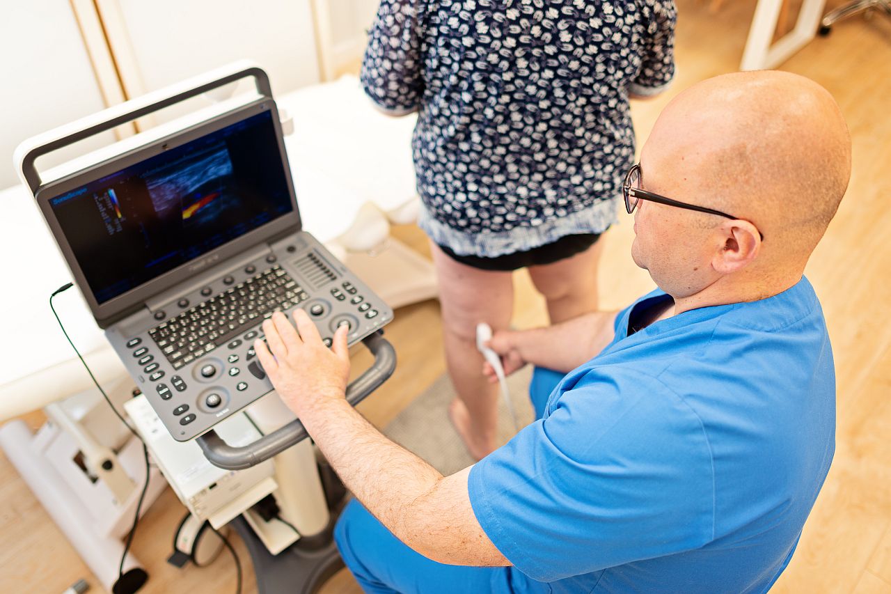
Doppler ultrasound examination
It’s worth checking the condition of your blood vessels before going on vacation. Based on the examination of color flow Doppler ultrasound, a specialist can accurately determine the condition of the superficial veins, deep veins and arteries. By following the doctor’s instructions, you can avoid many threatening complications and enjoy your vacation without worrying.
Author: VenoMedica
All rights reserved.
Author: venomedica
Date: 2019-06-04
5 most common myths about varicose veins
One of the most common vein diseases is called Chronic Venous Disease (CVD). Despite many studies, some aspects have still not been fully explained, and unfortunately, the knowledge of CVD disease is full of unreal information. Reasons as delaying treatment, unproven methods or incorrect recommendations are sadly leading to serious complications. With careful treatment however, you can reduce the risk of painful side effects.
Myth 1: Varicose veins are treated with drugs
Contrary to the popular belief, the so-called “drugs for Varicose veins” are most often normal dietary supplements, and can only be of secondary importance in the treatment of chronic venous insufficiency. They are not replacing basic methods of treatment such as surgical treatment or compression therapy. Such drugs are not the cause of disappearance of varicose veins nor the vascular spider veins. They will only expose the patient to unnecessary costs without reducing the risk of venous insufficiency complications.
Myth 2: Avoid any physical activity
It is proven that one couldn’t be more mistaken. It is the total opposite. Our calf-muscles work like a pump. When the muscles contract, they squeeze the deep veins in the legs together and transport the blood further, which stops the blood pooling in the legs so our legs and feet do not swell. The risk of developing thrombosis is then also being reduced. Therefore, activies such as hiking, climbing stairs or cycling keep the muscle pump fit and ensure good circulation to the legs.
Myth 3: Venous ulcers should be cured before any surgical treatment for varicose veins
This is a frequently repeated myth. Venous ulcers are chronic, non-healing wounds caused by advanced venous insufficiency accompanied by swelling of the subcutaneous tissue and recurrent inflammation. The basic condition for the wound to heal is the elimination of both the swelling and inflammation. The so-called ”conservative” treatment is usually difficult and expensive – it requires wearing of compression bandages or stockings for many months, which is a long time of distress for many patients. Current methods of closing inefficient veins, such as laser ablation (EVLT) or sclerotherapy, can be carried out even despite an open wound. Thanks to the combination of both treatment method and the appropriate dressing method, it is possible to significantly reduce pain and reduce the healing time to only several weeks instead of months.
Myth 4: Varicose veins should not be treated in summer
Summer months are a period of increased risk of complications of varicose vein disease. This is due to several factors. First of all, high temperature causes the expansion of superficial veins, which in turn leads to increasing of blood stasis in varicose veins. Secondly, the heat is no good for wearing tight compression stockings which most often makes the patients to give up. This can lead to inflammation of the subcutaneous tissue, venous ulceration or even thrombosis. Considering modern varicose veins treatment methods that are minimally invasive, it is often safer to undergo a surgery during summertime not exposing oneself to any complications that might instead ruin the vacation for the whole family. More in the article: Summer – the time to treat varicose veins
Myth 5: It is forbidden to travel by plane after a varicose vein surgery
There are no contraindications for traveling by plane even the second day after varicose vein surgery, provided it was performed using one of the minimally invasive methods. The patient after such a procedure is most often safely protected by compression products and anticoagulants, which is why travelling by plane itself does not increase the risk of complications after the treatment. This way, it is possible for people from abroad to consider coming to us for a treatment.
Author: VenoMedica
All rights reserved.
Author: venomedica
Date: 2019-05-27
Pilonidal Sinus Laser Treatment (SiLaT)
The pilonidal cyst is a chronic and disruptive condition of the intergluteal cleft area. This disease is 3-4 times more common in men, especially with a dark complexion, usually in the age range of 20-40. Factors such as excessive sweating, deep intergluteal cleft and dense body hair in this area, can contribute to the formation of a pilonidal cyst. Due to the long and difficult healing process, and the risk of recurrence, patients often struggle with this condition for many years, experiencing discomfort and embarrassment.
Pilonidal cyst: Causes
The exact causes of pilonidal cysts are still unclear. The most popular theory is that the pilonidal cyst forms as a result of hair trapped under the skin, caused by the forces acting during sitting down and standing up, as well as individual skin sensitivity. The condition usually manifests itself in the Sacrococcygeal area, as a lump filled with pus and hair. In young children, the causes can be different and be linked to organogenesis.
A painful problem
The location of pilonidal cysts often leads to infections, which cause forming of abscess. A painful lump will appear in the intergluteal cleft area, accompanied by redness of the skin, hard inflammatory infiltrate and often fever. There might foul-smelling pus or blood-tinged pus leaking out of the abscess. The untreated disease leads to reoccurring inflammations and infections, that often require surgical intervention.
Determining eligibility for treatment
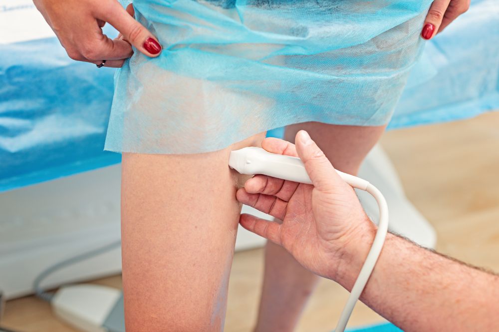
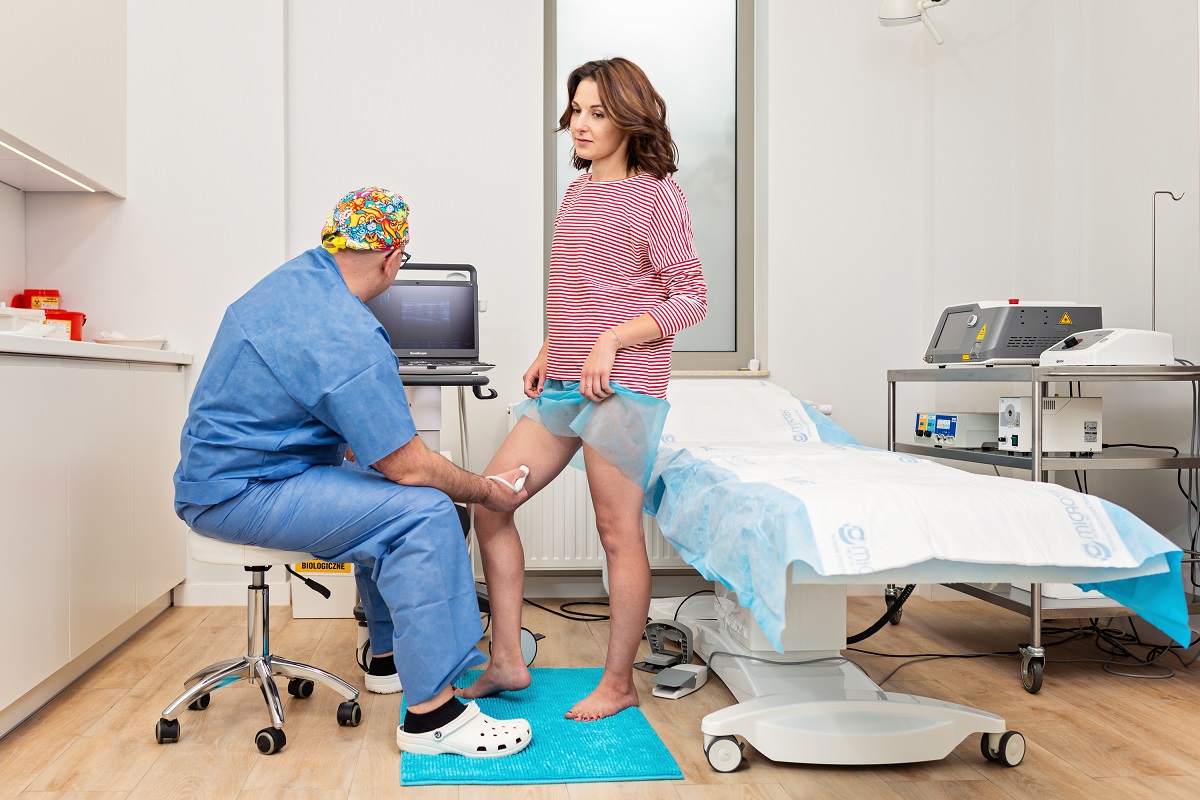
During the medical consultation, the doctor, based on the patient’s symptoms and the ultrasound scan, will assess the lesion and establish the final diagnosis. If the diagnosis confirms the pilonidal cyst, the doctor will assess the patient’s general health and decide about conducting a procedure, and which method is best for you.
Pilonidal cyst: Treatment
Typical treatment involves a radical removal of the cyst using different surgical techniques – from a simple removal to skin grafting. Classic surgeries are performed in the operating room under spinal or general anaesthesia followed by 2-3 days of hospital stay. Given the area of the body, where the surgery is performed, the infection rate is so high, that some surgeons tend not to close the wound with sutures and heal them by leaving the wounds open and allowing for granulation tissue to form. In such cases, the healing process can take up to a couple of months!
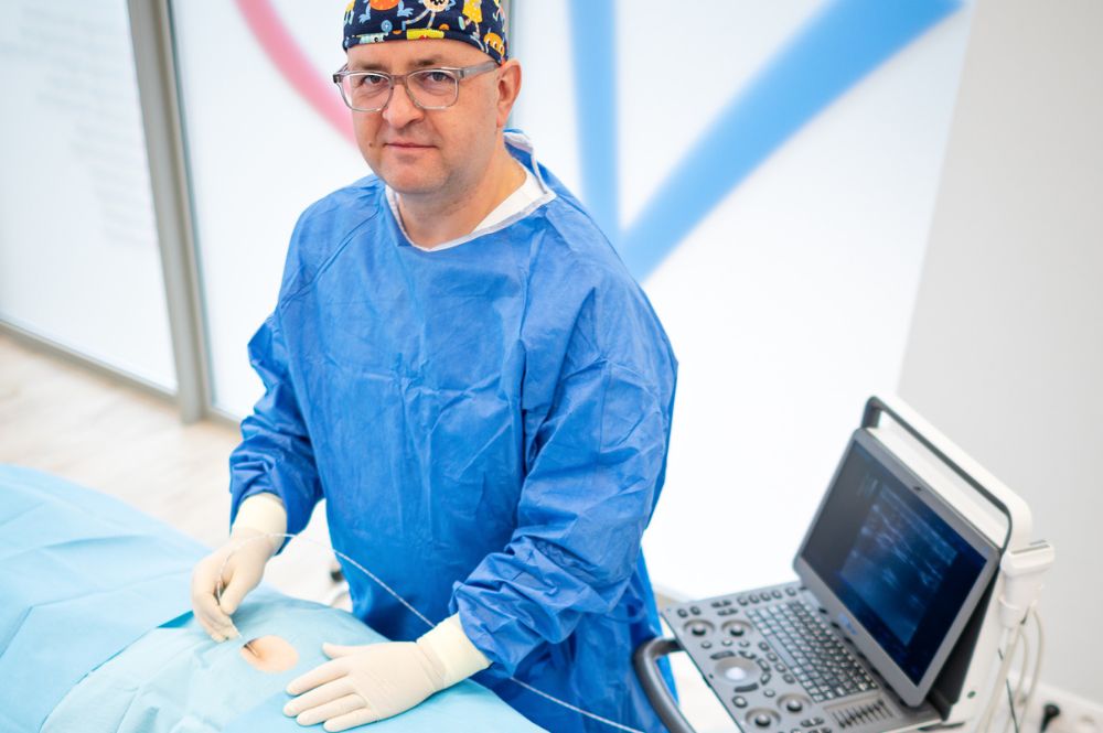
SiLaT laser treatment of pilonidal cyst
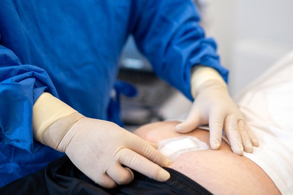
The latest and least invasive treatment method for a pilonidal cyst is laser ablation (SiLaT), using single-use radial fibres emitting 1470nm wavelength laser. After an incision is made in the abscess, it is drained and subsequently, upon inserting the laser fibre, the ablation of the cyst is performed. The procedure is performed under the local anaesthesia, on an outpatient basis, so the patient can go home directly after the procedure. The pain following the surgery is minor and the abscess incision wound heals within a couple of weeks. The effectiveness of the Pilonidal Sinus Laser Treatment (SiLaT) is very high and exceeds the effectiveness of most of the common surgical techniques while achieving a very good cosmetic result.
Author: VenoMedica
All rights reserved.
Author: venomedica
Date: 2019-03-14
Varicose veins and leg swelling – diagnosis and treatment of chronic venous insufficiency
Chronic venous insufficiency is a disease that is caused by impeding blood flow from the venous system. It may be caused by impaired valve function in either superficial veins (varicose vein disease) or deep veins (venous thrombosis). Venous insufficiency is caused by stagnation of blood in the venous system, which increases the blood pressure in the veins and leads to their significant dilatation over time. Dilated veins visible on legs are called varicose veins.
Swelling of the legs
Varicose veins are initially treated as a cosmetic problem. As a result of untreated disease, most often the area of sprain ankles appear swollen. Initially the swelling intensifies in the evening and tend to disappear after a night spent in a lying position. Over time however, they may become permanent not disappearing at all. They are caused by fluid penetrating from vessels filled with venous blood into the subcutaneous tissue. A characteristic symptom of swelling is a hole which remains in the skin when pressing down with a finger. Due to the plasticity of the swollen subcutaneous tissue, the swollen area is often described as “doughy”.
Inflammation and ulceration
The penetration of inflammatory cells into the fluid spilled over onto subcutaneous tissues, is causing skin inflammation. Its characteristic symptom are sclerosis and redness of both the skin and subcutaneous tissue, and its vivid soreness and increased skin temperature. The inflammation is usually located on the shins – near the ankles and on the front surface of the shin, bringing about problems related to skin allergy. A rash and numerous bubbles come out containing raw fluid causing very annoying itching. Patients scratch so hard that it comes to visible skin damage and abundant drainage of the serous fluid.
This mechanical damage to the inflamed skin can lead to ulceration, that is deep, non-healing wounds. Wounds are often infected, purulent-serous secretions causing constant pain and their healing time is long and difficult. Ulcers also have a tendency to relapse. Blood stasis in varicose veins initiates often venous thrombosis. A characteristic symptom is the hardening, redness and soreness of the skin in the vein area. Most often varicose vein thrombosis is local, however, in some cases it may pass into deep veins, which poses a threat to the patient’s life. The inflammation is giving way to brown skin discoloration, which very often persists for many years. After the healing period, unfortunately the ulcers leave extensive scars.
Advanced venous insufficiency may impair the efficiency and comfort of a patient’s life for many months or even years ahead. Lower limbs’ disability caused by venous diseases may intensify the formation of degenerative changes in limb joints as well as the spine. Early treatment gives excellent results, both therapeutic and cosmetic. Non-invasive treatment of venous insufficiency involves wearing properly selected compression products (knee socks or graduated compression stockings). Venous ulcers should be cleaned using individually selected dressings and preparations, under the supervision of experienced medical and nursing staff.
Diagnostics
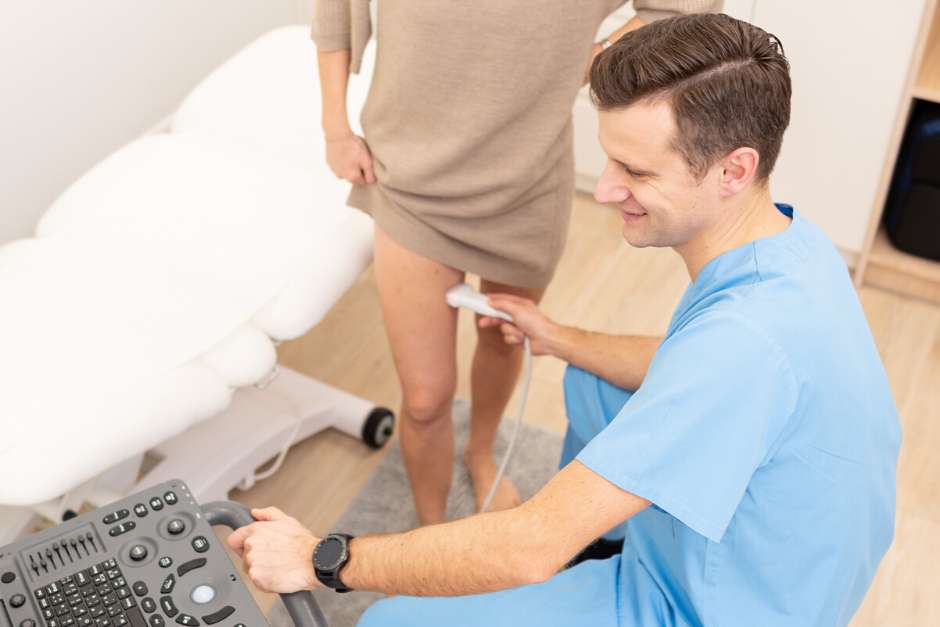
In the diagnosis and differentiation of venous diseases, the most important examination is Doppler ultrasound used to assess blood flow in the vessels. Based on the Doppler duplex ultrasound examination, the patient can be qualified for surgical treatment. If the cause of venous insufficiency is damage to the vein valves in the superficial system, then in order to reduce reflux, (i.e. reverse flow causing an increase in venous blood pressure) the damaged veins should be excluded from the circulation by closing them.
Currently the “gold standard” around the world are minimally invasive procedures performed in an outpatient procedure, which means that the patient can leave the medical center immediately after the procedure.
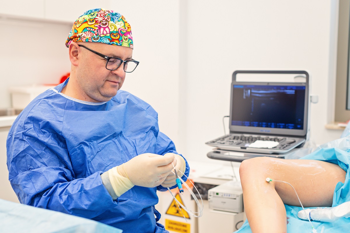
The most effective and safest methods include laser procedures performed under ultrasonography under local anesthesia (using a 1470 nm intravenous laser). The laser fiber is inserted into the vein through a small skin puncture. The energy from the laser light closes the diseased vein and blocks the blood supply to varicose veins. RF treatment is performed in a similar way, where the source of thermal energy is radio frequency current.
The mechanical-chemical methods are becoming very popular these days. It is a routine of mechanical destruction of the vein in order to damage the endothelium of the bulging vein by using a rotating catheter (e.g. ClariVein technology). The procedure is performed using a sclerosing agent leading to permanent vein occlusion. The procedure does not require anesthesia, thus the patient doesn’t feel any discomfort associated with skin puncture. Additionally, the procedures take less than 30 minutes. Sclerotherapy is very efficient when closing winding veins, small varicose veins and spider veins. The procedure uses a liquid sclerosant, usually administered as foam. Chemically, it is a detergent that causes irritation of the vascular endothelium, which leads to clotting of the vein and then after a few or even several weeks, it further leads to vein fibrosis then beginning to disappear. Sclerosants can also be used to close damaged veins. With those you can close both large venous trunks as well as small varicose veins.
Author: VenoMedica
All rights reserved.